Hip Muscles Diagram Labeled : 208 Vastus Lateralis Stock Photos Pictures Royalty Free Images : Human muscle system, the muscles of the human body that work the skeletal system, that are under voluntary control, and that are concerned with movement, posture, and balance.
Hip Muscles Diagram Labeled : 208 Vastus Lateralis Stock Photos Pictures Royalty Free Images : Human muscle system, the muscles of the human body that work the skeletal system, that are under voluntary control, and that are concerned with movement, posture, and balance.. The muscle of the arm is divided by a fascial layer separating the muscles into two osteofascial compartments: Human muscle diagram muscular system sketch at paintingvalley explore collection of. In this article we describe the hip and thigh muscles. Extension and rotation of the hip origin: Everyone should list the structures within muscle.
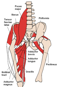
Most will label a diagram of muscle with its. Muscular compartment, bones (tibia, fibula) and muscles. Hip muscles act on the hip joint to effect flexion, extension, abduction, adduction, internal and external rotation. They are among the largest and strongest muscle in the body and are. Female hip and leg muscles labeled posterior view, 3d rendering.
Muscles Of The Hip Wikipedia from upload.wikimedia.org In human anatomy, the muscles of the hip joint are those muscles that cause movement in the hip. Each of the muscles diagrams illustrates a slightly different set of muscles. Human anatomy diagrams show internal organs, cells, systems, conditions. Human muscle diagram muscular system sketch at paintingvalley explore collection of. Human anatomy diagrams show internal organs, cells, systems, conditions, symptoms and sickness information and/or tips for healthy living. Learn vocabulary, terms and more with flashcards, games and other study tools. Learn vocabulary terms and more with flashcards games and other study tools. This diagram with labels depicts and explains the details of hip muscles diagram.
From physical best activity guide:
The four groups are the anterior group, the posterior group, adductor group, and finally the abductor group. The muscle of the arm is divided by a fascial layer separating the muscles into two osteofascial compartments: Most will label a diagram of muscle with its. Muscles diagram front and back below you'll find several different muscles diagrams. Their main function is contractibility. In this article we describe the hip and thigh muscles. Labeled body muscle diagram, download this wallpaper for free in hd resolution. Free access cross sectional anatomy of the hip : Don't forget to share this picture with others via facebook, twitter, pinterest or other social medias! Hip muscles are a group of seventeen to twenty skeletal muscles that provide a broad range of motion in the ball and socket joint of the hip. Back muscle diagram labeled best 15 bright human. Want to learn more about it? This hd wallpaper labeled body muscle diagram has viewed by 793 users.
Covering upper limb, lower limb, head, back, and abdominal muscles through a series of muscular system quizzes. Human anatomy diagrams show internal organs, cells, systems, conditions. Click on the labels below to find out more about your muscles. Cross section of the leg : Posted on january 20, 2015 by admin.
Gcse Pe Label Blank Muscles With Key Words Edexcel Worksheet Teaching Resources from dryuc24b85zbr.cloudfront.net It forms the medial wall of the femoral triangle. Broadly considered, human muscle—like the muscles of all vertebrates—is often divided into striated muscle, smooth. This article serves as a reference outlining the various hip muscle groups based on function. Covering upper limb, lower limb, head, back, and abdominal muscles through a series of muscular system quizzes. Posted on january 20, 2015 by admin. Hip muscles anatomy hip anatomy human body anatomy muscle anatomy leg muscles diagram muscle diagram lower. Everyone should list the structures within muscle. These muscles can be grouped based upon their location and function.
Want to learn more about it?
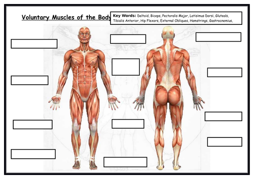
It forms the medial wall of the femoral triangle. Axial slice of mri with all anatomical structures labeled. Learn vocabulary terms and more with flashcards games and other study tools. The muscle of the arm is divided by a fascial layer separating the muscles into two osteofascial compartments: Click on the labels below to find out more about your muscles. The many muscles of the hip provide movement, strength, and stability to the hip joint and the bones of the hip and thigh. The anterior and the posterior compartments of the arm. Muscular compartment, bones (tibia, fibula) and muscles. An easy and convenient way to make label is to generate some ideas first. They are among the largest and strongest muscle in the body and are. Back muscle diagram labeled best 15 bright human. Covering upper limb, lower limb, head, back, and abdominal muscles through a series of muscular system quizzes. Label the major muscles of the body.
Their main function is contractibility. Label the major muscles of the body. Hip muscles are a group of seventeen to twenty skeletal muscles that provide a broad range of motion in the ball and socket joint of the hip. Most modern anatomists define 17 of these muscles, although some additional muscles may sometimes be considered. Want to learn more about it?
Muscle Lab 23 Figure 23 1 Muscles Of The Anterior Right Hip And Thigh Diagram Quizlet from o.quizlet.com Adductor tubercle on distal femur, entirety of linea aspera a: Everyone should list the structures within muscle. Click on the labels below to find out more about your muscles. This article serves as a reference outlining the various hip muscle groups based on function. Smartdraw includes 1000s of professional healthcare and anatomy chart templates that you can modify and make your own. Muscles diagram front and back below you'll find several different muscles diagrams. Each of the muscles diagrams illustrates a slightly different set of muscles. Extension and rotation of the hip origin:
It forms the medial wall of the femoral triangle.
This diagram depicts muscle labeled diagram. A complete list of muscular system quizzes; Anatomical diagram showing a front view of muscles in the human body. Human muscle diagram muscular system sketch at paintingvalley explore collection of. Posted on january 20, 2015 by admin. Label the major muscles of the body. Axial slice of mri with all anatomical structures labeled. Hip ad, extension adductor head: Muscles, connected to bones or muscles that act on the lower limb cause movement at the hip, knee and foot joints. In human anatomy, the muscles of the hip joint are those muscles that cause movement in the hip. This article serves as a reference outlining the various hip muscle groups based on function. Click on the labels below to find out more about your muscles. Hip muscles are a group of seventeen to twenty skeletal muscles that provide a broad range of motion in the ball and socket joint of the hip.
Everyone should list the structures within muscle hip muscles diagram. Human muscle system, the muscles of the human body that work the skeletal system, that are under voluntary control, and that are concerned with movement, posture, and balance.


0 Comments